Autologous PRP Injection into the Anterior Vaginal Wall for Female Sexual Health and Function
- 6 days ago
- 5 min read
Background Autologous platelet-rich plasma (PRP) has emerged as a regenerative biologic intervention in female sexual medicine, targeting stromal, vascular, and neurogenic components of genital tissue. One proposed application is submucosal injection into the distal anterior vaginal wall to improve sexual function, particularly orgasmic response and arousal.
Introduction
Female sexual dysfunction (FSD) is a multifactorial condition encompassing disturbances in desire, arousal, orgasm, and sexual satisfaction, often associated with distress and impaired quality of life. Current management strategies—pharmacologic (e.g., flibanserin, bremelanotide), hormonal therapy, psychosexual therapy, and energy-based devices—demonstrate variable efficacy and limited response in a substantial subset of patients.
Regenerative medicine approaches, particularly platelet-rich plasma (PRP), have emerged as a biologically plausible intervention aimed at enhancing tissue vascularity, neurogenesis, and extracellular matrix remodeling within genital tissues.
Biological Rationale and Mechanism of Action
Platelet-rich plasma (PRP) functions as an autologous biologic delivery system that transiently shifts the local tissue microenvironment from a quiescent, maintenance state toward a pro-regenerative, pro-angiogenic, and pro-remodeling signaling state. In genital tissue—particularly the distal anterior vaginal wall—this is relevant because the region is richly vascular, innervated (pudendal and pelvic plexus contributions), and hormonally responsive, yet susceptible to atrophic, ischemic, and neurosensory decline in conditions associated with female sexual dysfunction (FSD).
Mechanistically, PRP effects are mediated less by “cell replacement” and more by paracrine signaling cascades initiated by platelet degranulation following activation (endogenous thrombin exposure or exogenous CaCl₂).
1. Platelet Activation and Degranulation Biology Upon injection into tissue, platelets undergo:
Adhesion to extracellular matrix proteins (collagen types I/III, fibronectin)
Shape change and activation via integrin signaling
Degranulation of alpha granules
Alpha granules release:
Growth factors (PDGF, TGF-β, VEGF, IGF-1, EGF)
Chemokines (e.g., PF4/CXCL4, CCL5/RANTES)
Adhesion molecules (fibrinogen, vitronectin, fibronectin)
This produces a temporary cytokine-rich microenvironment lasting hours to days, which then initiates longer transcriptional and remodeling effects lasting weeks to months.
2. Growth Factor–Specific Mechanistic Pathways
Growth Factor | Primary Receptors / Pathways | Core Biological Effects | Vaginal Tissue Effects | Functional / Clinical Implications |
PDGF | PDGFR-α/β → MAPK/ERK, PI3K/AKT | Fibroblast proliferation, survival signaling, collagen gene upregulation (COL1A1, COL3A1) | ↑ Stromal fibroblast density; ↑ ECM turnover; improved structural integrity | Increased tissue turgor and mechanical responsiveness; potential enhancement of deformation-based sensory feedback |
TGF-β | TGF-βR → SMAD2/3 | Fibroblast → myofibroblast differentiation; ↑ type I collagen; MMP regulation; immunomodulation | Mucosal thickening; epithelial–stromal stabilization; wound-healing–like remodeling | Structural reinforcement; but excessive signaling may lead to fibrotic stiffening (dose-dependent risk) |
VEGF | VEGFR-2 (KDR/Flk-1) | Angiogenesis; endothelial proliferation; NO/eNOS activation; vascular permeability | ↑ Microvascular density; improved perfusion; capillary remodeling | Enhanced genital blood flow responsiveness, lubrication via transudation, improved oxygenation and arousal-related engorgement |
EGF | EGFR (ErbB1) → MAPK/ERK, PI3K/AKT | Epithelial proliferation, migration, survival | Increased epithelial turnover; improved stratification; mucosal barrier reinforcement | Improved lubrication, reduced microtrauma, enhanced epithelial sensory interface integrity |
IGF-1 | IGF-1R → PI3K/AKT, MAPK | Neurotrophic support; Schwann cell activation; axonal sprouting; anti-apoptosis | Supports peripheral nerve maintenance and repair in genital sensory fields | Potential enhancement of afferent sensitivity, orgasmic signaling efficiency, and recovery from neuropraxic or atrophic states |
3. Integrated Tissue-Level Effects in the Anterior Vaginal Wall
When combined, these growth factors produce a coordinated regenerative cascade:
Phase 1: Acute signaling (hours–days)
Platelet degranulation
Chemotaxis of macrophages and fibroblasts
Local inflammatory signaling (controlled, transient)
Phase 2: Proliferative phase (days–weeks)
Fibroblast activation (PDGF, TGF-β)
Endothelial proliferation (VEGF)
Epithelial regeneration (EGF)
Phase 3: Remodeling phase (weeks–months)
ECM reorganization (collagen maturation)
Increased vascular density
Nerve fiber stabilization and potential sprouting (IGF-1-mediated support)
4. Neurovascular Coupling and Sexual Function Correlates
Female sexual response is heavily dependent on neurovascular integration, particularly:
Pudendal nerve afferents (somatic sensation)
Pelvic plexus autonomic input (vascular engorgement)
Spinal reflex arcs (S2–S4 segments)
PRP may influence this system indirectly via:
Improved vascular supply
VEGF-mediated angiogenesis increases perfusion, improving oxygen delivery to nerve endings.
Enhanced nerve microenvironment
IGF-1 supports Schwann cell activity and axonal maintenance.
Improved mechanotransduction Tissue elasticity changes may enhance stimulation of mechanosensitive ion channels (Piezo channels) involved in sexual sensation.
5. Why the Lower Anterior Vaginal Wall?
This anatomical region is selected due to:
Dense periurethral vascular plexus
High innervation density (somatic + autonomic convergence)
Proximity to clitoral crura neurovascular network
Mechanical stimulation during penetration (high functional relevance)
It is therefore a high-yield target zone for neurovascular modulation therapies, analogous in concept (not structure) to intracavernosal targeting in male sexual medicine.


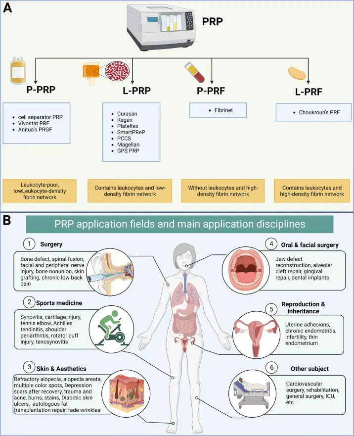
Technical Considerations: PRP Preparation and Injection Protocol
PRP Preparation Variables (Critical for reproducibility)
Blood draw: 10–60 mL autologous peripheral blood
Centrifugation: Typically 1–2 spin systems
Platelet concentration target: 3–5× baseline
Leukocyte content: Leukocyte-rich vs leukocyte-poor (protocol dependent)
Activation: Calcium chloride or thrombin (not universally used)
Clinical outcomes in genital applications are highly sensitive to PRP composition, especially leukocyte concentration and platelet yield.
Injection Technique (Anterior Vaginal Wall)
Category | Specification | Details |
Anatomical Target | Distal anterior vaginal wall | Distal one-third of anterior vaginal wall |
Regional anatomy correlation | Periurethral and paraurethral tissue complex; overlaps with proposed “G-spot” region (hypothesis-dependent anatomy) | |
Patient Positioning | Lithotomy position | Standard gynecologic examination/procedural positioning |
Preparation | Aseptic technique | Antiseptic solution applied to operative field prior to injection |
Instrumentation | Needle gauge | 21–27G needle |
Delivery system | Syringe (typically 1–3 mL per injection site) | |
Injection Technique | Tissue plane | Submucosal injection plane |
Distribution method | Multiple small-volume aliquots along distal anterior vaginal wall | |
Safety precaution | Aspiration prior to injection to reduce risk of intravascular delivery | |
Session Protocol | Treatment frequency | 3–4 sessions |
Interval between sessions | Approximately 3–4 weeks (study-dependent variability) |
Post-procedure Care
Avoid sexual intercourse for 48–72 hours (varies by protocol)
Monitor for spotting, mild edema, or transient discomfort
No routine antibiotics unless indicated
Safety Profile
Across published regenerative gynecology literature, PRP demonstrates a favorable safety profile due to autologous origin.
Reported adverse effects:
Mild pain at injection site
Transient edema or erythema
Spotting or minor bleeding
Rare infection (typically technique-related
Comparative Positioning in Female Sexual Medicine
PRP is conceptually positioned within regenerative sexual medicine, alongside:
CO₂ and Er:YAG vaginal lasers
Radiofrequency tissue remodeling
Hyaluronic acid-based vaginal fillers (investigational)
Hormonal optimization strategies
Unlike energy-based devices, PRP directly introduces autologous bioactive mediators, potentially shifting tissue biology rather than inducing controlled thermal injury.
Conclusion Autologous PRP injection into the lower anterior vaginal wall represents a promising regenerative approach for female sexual dysfunction, particularly in orgasmic disorder and sexual satisfaction deficits. The study by Sukgen et al. demonstrates statistically and clinically meaningful improvements in FSFI and orgasmic function, with high patient-reported satisfaction and minimal adverse effects.
Reference:
Sukgen, G., Kaya, A. E., Karagün, E., & Çalışkan, E. (Year not provided in abstract). Platelet-rich plasma administration to the lower anterior vaginal wall to improve female sexuality satisfaction. [Study details from provided abstract].
Discover the Korean approach to Non-surgical Female Rejuvenation from O-shots to Vagina Rejuvenation with Neurotoxins:
IFAAS Mini Fellowship (Hands-On)
Non-surgical Aesthetic Gynaecology: Injectables & Light Based Devices

16 August, 2026 - Seoul, South Korea - [Register Now]
More Upcoming Global Events
Comments