


Chin and Jawline Filler: A Precision-Based Approach to Natural Lower-Face Definition
Introduction
The most convincing chin and jawline treatments rarely begin with the syringe.
They begin with a diagnosis.
A patient may describe a “weak jawline,” an overly prominent nose, jowling, or even a double chin. Yet the underlying issue may be insufficient chin projection, inadequate mandibular support, soft-tissue distribution, asymmetry, or a combination of these factors. Treating the complaint in isolation can produce excessive volume without restoring facial balance.
A prospective case series by Alcantara and colleagues explores a more structured approach: individualized lower-face assessment, anatomically selected injection planes, high-elastic-modulus hyaluronic acid filler, and ultrasonographic and three-dimensional imaging evaluation.
Although the study is small, its findings offer several useful lessons for clinicians seeking natural results while limiting unnecessary product use.
Anatomical Considerations for a Safe Procedure
Safe chin and jawline augmentation requires more than identifying the mandibular border. The lower face contains variable vascular, neural, muscular, and glandular structures, and their relationships may change with age, facial morphology, previous procedures, and soft-tissue thickness.
Several structures deserve particular attention:
Facial artery and vein: The facial artery typically crosses the inferior mandibular border near the antegonial notch before continuing toward the oral commissure. Its course and depth are variable, however, making landmark-based prediction imperfect. Intravascular injection may result in vascular occlusion, ischemia, and tissue necrosis.
Mental foramen and mental neurovascular bundle: Usually located near the premolar region, the mental foramen transmits the mental nerve and accompanying vessels. Injection within or close to this area may cause pain, paresthesia, dysesthesia, neuropraxia, or vascular injury.
Marginal mandibular nerve: This branch of the facial nerve may travel close to or below the mandibular border. It can become particularly vulnerable in patients with limited subcutaneous tissue or age-related soft-tissue changes. Injury may produce temporary or, rarely, persistent lower-lip weakness and asymmetry.
Parotid gland and duct: Posterior jawline and mandibular-angle treatment must account for the parotid gland, while more anterior injections should consider the course of the parotid duct. Incorrect placement may contribute to glandular irritation, inflammation, or functional injury.
Superficial temporal artery: Posterior treatment near the preauricular region requires caution because this vessel is commonly located anterior to the tragus. The study positioned posterior access approximately one fingertip, or around 1 cm, anterior to the tragus; nevertheless, individual vascular anatomy should be assessed rather than assumed.
Submental structures: Excessive or poorly positioned filler near the anterior digastric muscles and submental space may blunt the cervicomental angle, create heaviness, or interfere with natural lower-face movement.
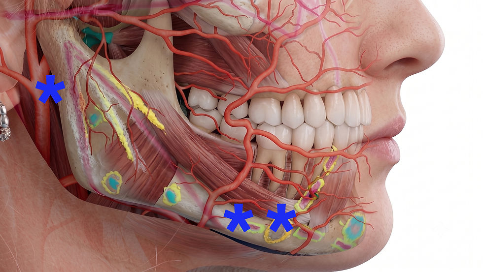
A layered anatomical assessment should therefore precede treatment. Palpation and surface markings can help orient the injector, but they cannot reliably identify every anatomical variation. When available—and when used by a properly trained clinician—high-frequency ultrasound enables direct evaluation of vessels, tissue planes, pre-existing filler, and areas of altered anatomy.
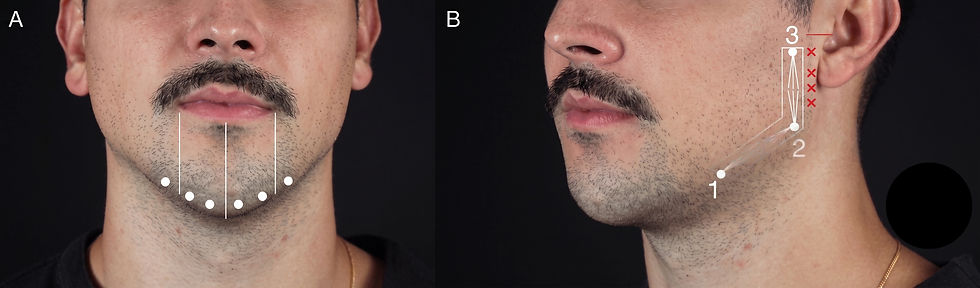
In the reported protocol, ultrasound was used during treatment to confirm injection-plane accuracy and again at follow-up to evaluate filler position and soft-tissue response. The investigators standardized the examination at the mandibular border, midline menton, and mandibular-notch region, using a positioning template to improve comparison between visits.
Risk reduction should incorporate multiple safeguards:
Detailed anatomical and medical assessment
Identification of previous filler, surgery, trauma, or scarring
Conservative, incremental product deposition
Appropriate selection of needle or cannula
Deliberate control of injection depth and direction
Slow injection with minimal pressure
Continuous observation for pain, blanching, livedoid change, or altered capillary refill
Ultrasound guidance when available and clinically appropriate
Immediate access to a complication-management protocol and hyaluronidase when injecting hyaluronic acid filler
Ultimately, ultrasound can improve anatomical awareness, but technology does not make an injection intrinsically safe. Outcomes still depend on training, product knowledge, appropriate case selection, controlled technique, and the clinician’s ability to recognize and manage complications promptly.
Treatment Planning and Injection Technique
Successful lower-face contouring begins by defining the structural problem—not by deciding how many milliliters to inject.
Assessment should include frontal, oblique, profile, and dynamic views. The clinician should evaluate chin projection and height, pogonion and menton position, bigonial-to-bizygomatic proportions, gonial angle, prejowl depression, mandibular-border continuity, soft-tissue thickness, and submental contour. Dental relationships, occlusion, skeletal discrepancies, facial asymmetry, and prior aesthetic procedures may also influence the treatment plan.
The patient’s original complaint should be interpreted within this wider assessment. Apparent nasal prominence, early jowling, or submental fullness may be accentuated by inadequate chin projection or mandibular support. Recognizing these relationships can prevent treatment of the wrong anatomical target and reduce unnecessary product placement.
Cephalometric relationships and the golden ratio may provide useful reference points, but they should not be treated as universal prescriptions. In the study, these concepts were used qualitatively to guide treatment rather than as standardized numerical endpoints. Planning remained individualized according to facial structure, age, ethnicity, tissue distribution, and patient preference.


Selecting the Filler
The investigators used a high-elastic-modulus, or high-G′, hyaluronic acid filler. Products with greater resistance to deformation may be suitable for structurally demanding areas in which projection and shape retention are required.
Filler selection should nevertheless account for more than G′ alone. Cohesivity, viscosity, tissue integration, swelling behavior, reversibility, injection force, and the manufacturer’s approved indications should all be considered. The product selected for deep chin support may not be ideal for every superficial contouring requirement.
Chin Projection: Deep Structural Support
For chin augmentation, the study used needle-delivered boluses in the deep supraperiosteal plane. Placement close to bone was intended to provide structural projection and make efficient use of the high-G′ filler.
The injection pattern was adjusted to the desired morphology. Broader distribution across four to eight points was used when a wider, squarer chin was desired. A three-point pattern—with greater emphasis on the central point—was used to create narrower projection and a more tapered contour.
These patterns should be considered adaptable references rather than sex-based rules. Some patients prefer a stronger angular result, while others favor elongation, central projection, or softer transitions regardless of sex or gender identity.
Bolus size, number of points, and total volume should be determined by the severity of deficiency, tissue thickness, skeletal anatomy, product characteristics, and tolerance of the overlying soft-tissue envelope. Large boluses or excessive lateral placement may produce overprojection, widening, compression, irregularity, or distortion during animation. Jawline and Mandibular Angle: Subcutaneous Contouring
For the mandibular body and angle, the authors used a 22G blunt cannula in the subcutaneous plane. Filler was delivered through retrograde linear threads, with no more than 0.2 mL reported per line.
This approach was intended to create a smooth, visible contour while limiting the volume required to overcome deeper muscle and soft tissue. The cannula entry point and direction were adapted to the region being treated, with attention to the masseter, facial vessels, marginal mandibular nerve, parotid structures, and preauricular vasculature.
Subcutaneous placement is not automatically risk-free. Cannulas may still penetrate vessels, nerves, glands, or deeper planes, particularly when advanced with force or through scarred tissue. The instrument should therefore be moved gently, without assuming that reduced penetration risk equals elimination of risk.
Planning the Sequence
A useful treatment sequence is to establish central support before refining the lateral contour:
Correct clinically meaningful chin retrusion or vertical deficiency.
Reassess the profile, lower facial thirds, and cervicomental relationship.
Treat prejowl depressions when they interrupt mandibular continuity.
Refine the mandibular body and angle only to the extent required.
Reassess symmetry from multiple views after each incremental deposition.
This staged approach may prevent compensatory overfilling of the jawline when inadequate chin projection is the primary source of imbalance.
Volume Should Follow Anatomy
Patients in the case series received a total of 3-6 mL, depending on the number of regions treated. The objective was not to reach a predetermined volume but to place product where it would create the greatest structural effect.
Soft-tissue thickness can substantially influence the result. A thin tissue envelope may display projection from a relatively small volume but may also reveal superficial irregularities. Thicker tissue may conceal definition and require a different plane, product, or treatment strategy. Severe skeletal deficiency, substantial skin laxity, or significant submental adiposity may not be adequately corrected with filler alone.
Clinicians should therefore recognize when filler is unlikely to meet the patient’s goals. Orthognathic assessment, genioplasty, energy-based treatment, lipolysis, or surgical neck and lower-face procedures may be more appropriate in selected cases.
The desired endpoint is not maximal projection. It is a continuous mandibular contour that remains balanced at rest and during movement, with no visible transition between treated and untreated regions.
Shape Should Be Personalized—not Prescribed by Sex Alone
The study describes different reference patterns for traditionally masculine and feminine lower-face aesthetics.
The masculine treatment pattern emphasized:
A wider, squarer chin
Greater bigonial width
A more angular mandibular contour
A gonial angle referenced at approximately 90° to 110°
The feminine pattern emphasized:
A narrower or tapered chin
Subtle elongation
Softer mandibular transitions
Preservation of midface dominance relative to bigonial width
These patterns can be useful starting points, but they should not override individual preference. The authors appropriately note that desired masculine or feminine characteristics should be adapted to each patient, regardless of gender identity.
The clinical objective is not to impose a standardized facial ideal. It is to understand what combination of projection, width, length, and angularity will harmonize with the individual face.
Anatomy Remains the Foundation of Safety
Lower-face filler treatment involves several structures with important safety implications:
The facial artery and vein
The mental foramen and neurovascular bundle
The marginal mandibular branch of the facial nerve
The parotid gland and duct
The superficial temporal artery near posterior access points
The anterior digastric and submental spaces
The facial artery commonly crosses the mandibular border near the antegonial notch, but its course and depth can vary. The mental foramen must be considered when placing product near the premolar region. Posterior mandibular treatment requires awareness of the parotid structures and vessels near the preauricular region.
The study combined anatomical landmarking, controlled plane selection, cannula use in selected regions, aspiration before needle-based boluses, and ultrasonographic guidance.
Aspiration should be understood as only one component of a broader safety strategy. A negative aspiration result cannot reliably exclude vascular placement. It does not replace detailed anatomical knowledge, slow low-pressure injection, conservative deposition, continuous observation, appropriate instrument and plane selection, or readiness to recognize and manage vascular compromise.


What Ultrasound Adds
Ultrasound can help clinicians visualize soft-tissue layers, identify vessels, confirm product placement, and investigate suspected complications. In this study, an 11-MHz high-frequency linear transducer was used during treatment and follow-up.
The authors standardized imaging at three locations:
The mandibular border, approximately 1 cm anterior to the mandibular angle
The midline menton
The mandibular notch region, approximately 1 cm posterior to the oral commissure
A positioning template was used to improve consistency between visits.
This represents an important shift in aesthetic medicine: imaging can move treatment beyond landmark-based inference toward direct anatomical visualization. However, ultrasound is operator-dependent. Its value relies on adequate training, correct interpretation, and integration with—not replacement of—clinical anatomy.


Product Efficiency Is About Placement, Not Simply Low Volume
The study’s title and conclusions emphasize achieving natural results with relatively small product volumes. Yet “minimal volume” should be interpreted in context: patients received between 3 and 6 mL in total.
The more meaningful message is therefore not that every lower-face treatment requires very little filler. It is that carefully targeted placement may reduce the volume needed to achieve a defined structural objective.
Soft-tissue thickness is particularly relevant. A thin tissue envelope may allow a smaller amount of filler to create visible projection, while thicker tissue may require a different strategy. Conversely, superficial placement in thin tissues may increase the risk of visibility, irregularity, or palpability.
Product selection, tissue thickness, injection depth, and skeletal deficiency should therefore be considered together.
Important Limitations
The study provides a useful clinical framework, but several limitations restrict the strength of its conclusions:
The sample included only 10 patients.
All treatments were performed at a single center by one injector.
There was no control or comparator group.
Follow-up was limited to 90 days.
Ultrasound and 3D imaging were not reported uniformly for every participant.
Cephalometric and golden-ratio assessments were used qualitatively rather than as standardized endpoints.
Patient satisfaction was reported as a percentage, but the measurement instrument was not clearly described.
The authors reported industry involvement: the filler supplier provided the products and funded the publication fee, and one author disclosed consultancy fees from the supplier.
These factors do not invalidate the observations, but they mean the technique should be viewed as promising rather than definitively proven. Larger prospective studies with validated outcome scales, blinded evaluators, consistent imaging, and follow-up of at least 6 to 12 months are needed.
Practical Takeaways for Clinicians
This case series supports five practical principles:
Diagnose the structural cause of the concern. A complaint involving the nose, jowls, or submental area may originate partly from inadequate chin or mandibular support.
Plan in three dimensions. Frontal width, profile projection, vertical chin height, asymmetry, and the face-neck transition should be evaluated together.
Match rheology and plane to purpose. Structural projection and surface contouring have different mechanical and anatomical requirements.
Individualize the desired shape. Sex-associated aesthetic patterns may provide references, but patient preference and existing anatomy should direct treatment.
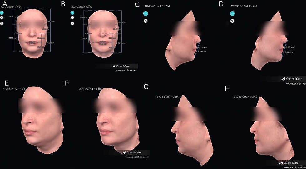
Use imaging thoughtfully. Ultrasound and 3D photography can strengthen planning and documentation, but neither replaces anatomical expertise or validated clinical outcome measures.
The Bottom Line
Natural chin and jawline enhancement is not simply a matter of adding volume along the mandibular border. It is an exercise in structural diagnosis, proportion, anatomical precision, and restraint.
This prospective case series suggests that a targeted combination of deep chin support and subcutaneous mandibular contouring can produce measurable short-term improvements with high patient satisfaction and few transient adverse effects. Ultrasound and 3D imaging add valuable objective information about injection depth, filler behavior, and facial change.
The findings are encouraging, but preliminary. The most defensible conclusion is that anatomically guided, individualized treatment may improve product efficiency and aesthetic coherence—not that one standardized protocol is appropriate for every patient.
For clinicians, the central message is simple: assess globally, inject purposefully, image when appropriate, and allow anatomy—not volume—to lead the treatment plan. Reference:
Alcantara A, Vassoler Guerrero Puccia É, Perrella de Rezende C, Kim JH, Viana R. Optimizing Dermal Filler for Chin and Jawline Definition as an Advanced Approach for Natural Results: A Prospective Case Series With Ultrasonographic and 3D Facial Imaging Evaluation. Cureus. 2025;17(12). doi:10.7759/cureus.99477.
Master advanced techniques in skin rejuvenation using injectables, lasers and next-generation biostimulators this December in Tokyo, Japan:
Mini Visiting Fellowship (Observation)
Japanese Tailored Aesthetic Injectables & Combination Therapies

17-18 December, 2026 - Tokyo, Japan - [Register Now]
More Upcoming Global Events