


Dual Cortical Tunneling Method for Endoscopic Forehead Lift
A clinical review of Kim and colleagues’ 2022 study
Introduction
Maintaining brow elevation after endoscopic forehead lifting remains a clinical challenge. In the study reviewed here, Kim and colleagues examined whether modifying cortical tunnel fixation could reduce postoperative brow relapse.
Endoscopic forehead lifting offers an approach to upper facial rejuvenation through limited incisions. Yet maintaining the lifted tissues remains a technical challenge. Even after adequate release, concentrated tension at a fixation point can allow sutures to gradually cut through soft tissue, contributing to recurrent brow descent.
In their 2022 study, Dual cortical tunneling method for endoscopic forehead lift, Kim and colleagues explored a modification designed to address this problem. Their approach distributes fixation across a broader tissue plane, offering useful insights into the relationship between anatomical release, mechanical support and postoperative stability.
This review follows the authors’ approach from anatomical release and patient assessment to fixation and 6 month outcomes. It focuses on the reported technique and the limits of the evidence.

Anatomical Foundations of Brow Position and Fixation
The study’s anatomical focus is the relationship between the brow depressor muscles, periorbital ligamentous attachments and the supporting layers incorporated into fixation.
The study highlights the importance of managing the brow depressors: the corrugator supercilii, depressor supercilii, procerus and medial portion of the orbicularis oculi. The authors emphasise that appropriate management of these muscles, together with release of the relevant periorbital ligaments, is a prerequisite for meaningful assessment of any fixation method.
Fixation supports repositioned tissue, while adequate release allows that tissue to reach its intended position. The periosteum and galea also play an important role in the authors’ technique. Incorporating these deeper supporting layers aims to distribute tension and stabilise the forehead flap while tissue adhesion develops.
Assessment must also account for frontalis compensation. Patients may recruit their forehead muscles to elevate the brows, potentially masking the resting position. To reduce this influence, the investigators measured brow position with patients’ eyes closed. Their approach reinforces the value of consistent assessment conditions when documenting preoperative anatomy and postoperative change.

Technique and Patient Consideration
Planning around the individual brow
Patients underwent preoperative photography and surgical marking while seated. The investigators assessed the medial and lateral brow separately, documenting both the initial elevation and subsequent relapse.
The authors particularly discuss medial brow elevation in Asian patients and caution that excessive vertical elevation laterally may accentuate an existing upward slant. This supports individualised planning rather than applying a uniform lifting pattern across patients.
From focal fixation to distributed support
The reported approach used five incisions: one central, two paramedian and two temporal. Cortical fixation was performed at the central and paramedian sites. Conventional single-tunnel fixation used one tunnel at each of these three sites. The dual-tunnel technique added a second tunnel at each site, creating six tunnels in total.
Caudal fixation incorporated tissue from the subcutaneous level through the periosteum, while cephalic fixation engaged the periosteum and galea. Together, these fixation points created a triangular supporting plane intended to reduce focal loading and the “cheese-wiring” effect associated with gradual tissue cut-through.


Variable | Total (n = 200) | Single tunnel (n = 100) | Dual tunnel (n = 100) | P-value |
Preoperative brow position (medial) | 16.81 ± 1.74 | 16.89 ± 1.97 | 16.72 ± 1.49 | 0.492 |
Preoperative brow position (lateral) | 19.41 ± 2.33 | 19.88 ± 2.44 | 18.93 ± 2.11 | 0.004 |
Elevated distance (medial) | 8.17 ± 0.96 | 8.36 ± 1.04 | 7.98 ± 0.84 | 0.006 |
Elevated distance (lateral) | 7.69 ± 1.42 | 7.78 ± 1.57 | 7.60 ± 1.24 | 0.370 |
Immediate postoperative brow position (medial) | 24.97 ± 1.71 | 25.25 ± 2.06 | 24.70 ± 1.22 | 0.024 |
Immediate postoperative brow position (lateral) | 27.10 ± 2.15 | 27.66 ± 2.28 | 26.53 ± 1.85 | < 0.001 |
6 months postoperative brow position (medial) | 22.94 ± 1.61 | 22.86 ± 1.88 | 23.01 ± 1.31 | 0.513 |
6 months postoperative brow position (lateral) | 24.38 ± 2.08 | 24.40 ± 2.26 | 24.37 ± 1.89 | 0.906 |
Relapsed distance (medial) | 2.04 ± 0.75 | 2.39 ± 0.83 | 1.69 ± 0.46 | < 0.001 |
Relapsed distance (lateral) | 2.71 ± 0.94 | 3.26 ± 0.91 | 2.17 ± 0.59 | < 0.001 |
Table 1. Serial measurement of the eyebrow position in terms of the distance between the lower margin of the eyebrow and the upper eyelid
Technical considerations for effective fixation
Dual tunneling required incisions approximately 2–3 mm longer and added around 5–15 minutes to operating time. The authors acknowledge a learning curve. The technique avoids a separate implanted fixation device, although sutures remain part of the construct.
The authors emphasise that appropriate management of the brow depressor muscles and release of periorbital ligaments are prerequisites, regardless of the fixation method selected. Their comparison therefore concerns fixation after these steps have been performed.
After Surgery and Results
The study included 100 patients aged 17–65 years, with 50 patients in each fixation group. Brow position was assessed before surgery, immediately afterwards and at six months. Both groups experienced postoperative descent, but the dual-tunnel group demonstrated significantly less relapse from the immediate postoperative position.
Smaller relapse did not translate into a statistically significant difference in absolute brow position between groups at six months. Baseline positions and aspects of the initial correction differed. These findings support improved maintenance of the immediate lift within this series, rather than proving a higher final brow position.
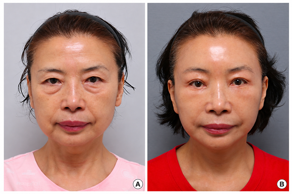
No significant hematoma, nerve damage, alopecia or infection was observed during six months of follow-up. No incision-site alopecia or unacceptable scarring was reported. These observations concern this study cohort; they do not establish the absence of risk. The paper does not provide a detailed postoperative recovery protocol or comparative outcomes beyond six months.
Conclusion In this series, dual cortical tunneling was associated with less medial and lateral brow relapse over six months than single-tunnel fixation. Absolute brow positions at six months were not significantly different. The authors’ findings support further consideration of distributed fixation after appropriate anatomical release. For surgeons studying this approach, the evidence highlights the importance of understanding both the fixation mechanics and how postoperative stability is measured.
Reference
Kim M, Lee N, Tark W, Lee WJ, Roh TS, Baek W. Dual cortical tunneling method for endoscopic forehead lift. Arch Aesthetic Plast Surg. 2022;28(3):85–89. https://doi.org/10.14730/aaps.2022.00444
Explore Korean endoscopic forehead and brow lift techniques through live surgery observation, focusing on minimal scarring and complication management. IFAAS Mini Fellowship (In-OR Observation)
Korean Advanced Facial Aesthetic Surgeries - Advanced Asian Blepharoplasty

11 March, 2027, Seoul, South Korea - [Register Now]