


Penile HA Filler: The Anatomy Behind the Result
Introduction
In penile HA filler enhancement, placement matters as much as volume.
A recent case report uses ultrasound to show where filler was placed within the penile shaft, how it was distributed and what changed at one month.
For clinicians, the result extends beyond a gain in circumference. Patient expectations, the target anatomical plane, even distribution and changes in contour over time all shape the assessment. This single case offers a close view of the technique; longer follow-up studies provide context for how results may evolve.
The anatomy: understanding the target plane
The penile shaft contains several distinct layers. Beneath the skin lies the dartos fascia; deeper still is Buck fascia, which surrounds the erectile structures. The dorsal neurovascular bundle and the urethra are key structures to consider when planning treatment.
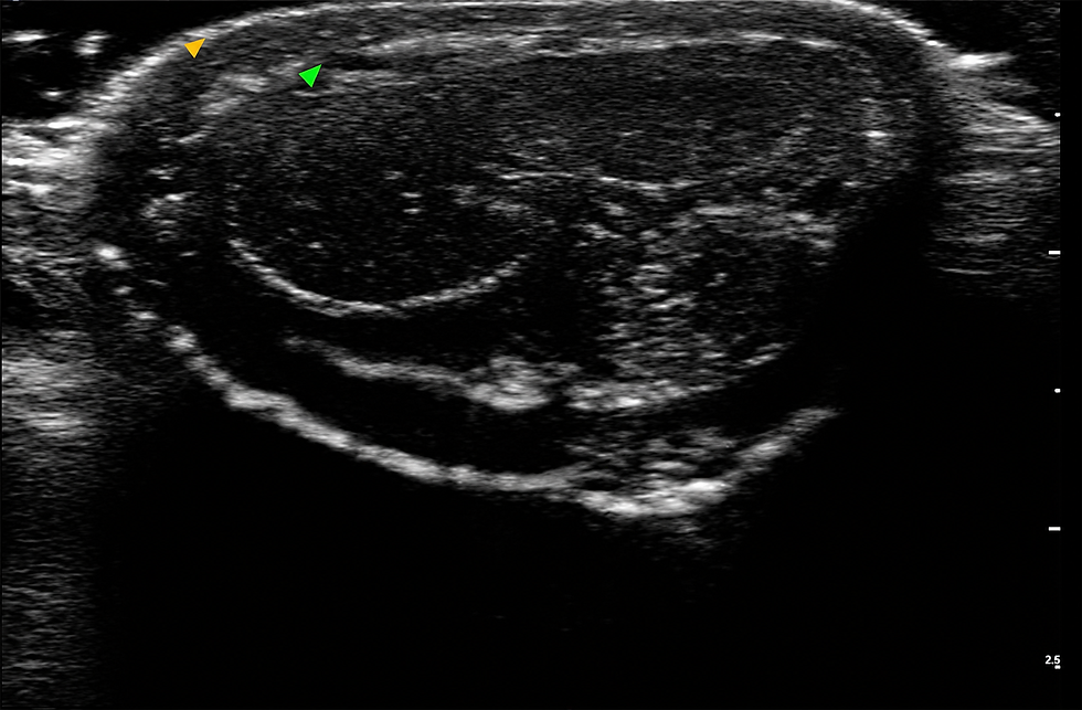
In a 2025 case report published in Plastic and Reconstructive Surgery – Global Open, the treating team placed HA fillers between the Dartos and Buck fascia. Ultrasound was used to visualise that anatomical plane and assess placement. This is the central educational feature of the paper: the images connect the visible change in shaft contour with the location of filler beneath the skin.
An improved external contour alone cannot confirm injection depth. Ultrasound provided additional anatomical documentation in this case, although one report cannot establish whether imaging improves safety or outcomes compared with other approaches.

Planning: the patient before the procedure
The case involved a 35-year-old man who felt that his penile girth was inadequate and said the concern affected his confidence. He had no previous genital surgery or penile augmentation. His medical history was otherwise unremarkable: the report notes no significant medical or psychiatric conditions, no regular medication use and no history of erectile dysfunction. It also reports normal endocrine function.
The assessment addressed why he wanted treatment, as well as his physical suitability. According to the case report, counselling did not identify penile dysmorphic disorder, and the team judged his expectations to be realistic. On examination, his BMI was 24.5 kg/m², with no excess pubic fat or protruding abdomen noted. The team found no contraindication in its assessment and proceeded with HA filler enhancement.
For clinicians, this part of the case illustrates the value of clarifying the patient’s concern before planning volume or technique. The report documents an aesthetic request, without describing a functional problem that the filler was intended to treat. A suitable result therefore needed to be discussed in terms of the patient’s appearance goals and realistic expectations, alongside the anatomical assessment.
How should the technique be adapted to the patient?
The ultrasound case used 5 mL of HA, a 22G cannula and two entry points at the penile base. Other published studies have used different volumes, cannulas and access patterns. These differences reflect distinct products, techniques and study populations; they have not been tested head to head to determine which combination is best for a particular patient.
Clinical consideration | What the literature reports | Question for treatment planning |
Baseline anatomy and contour | The ultrasound case placed filler between dartos and Buck fascia and used imaging to assess the plane. The 2026 study measured girth at the proximal, midshaft and distal shaft rather than relying on one measurement. | Where is additional volume sought, and how will placement and symmetry be assessed? |
Cannula and access | The ultrasound case used a 22G cannula and two basal entry points. A separate clinical trial used a different entry-point pattern. These are descriptions of study methods, not evidence that either approach suits a defined patient subgroup. | Can the intended plane and distribution be achieved while accounting for the patient’s anatomy and prior treatment? |
Initial volume | Published studies range from the 5 mL single-patient case to substantially larger volumes in clinical cohorts. The 2026 study deliberately adjusted initial volume and used later contour correction rather than treating the largest possible first-session increase as its objective. | What volume is appropriate for the planned contour, tissue characteristics and the patient’s expectations? The papers do not provide a universal calculation. |
Prior filler or existing irregularity | In the 2026 cohort, 12 of 20 patients had previous girth enhancement. A separate four-patient case series describes residual HA that required assessment and management before further augmentation. | Is existing material contributing to the current shape, and does it need evaluation before more filler is considered? |
Early follow-up | The ultrasound case reported a one-month outcome. The 2026 study distinguished smaller retouch procedures for contour irregularities from later reinjection for renewed augmentation. | How will contour and patient satisfaction be reassessed after the initial treatment period? |
The practical lesson is to plan for the patient’s anatomy and the desired contour, then assess the result over time. A cannula gauge, entry-point pattern or volume reported in one paper is a record of that team’s method—not a patient-selection algorithm or a substitute for clinical training.

Before and after: what was measured?
At one month, the patient’s flaccid midshaft circumference measured 13.0 cm, compared with 12.3 cm before treatment—an increase of 0.7 cm. The report describes patient satisfaction with the appearance and no major complication during its short follow-up.
That is a documented early result for one patient, not an expected result for all patients. It also cannot answer whether the measured increase or contour will persist for years. Those questions require larger groups and longer observation.
Longevity: what do longer studies show?
A 2026 prospective study of 20 patients followed penile girth measurements and patient-reported outcomes for 48 weeks after HA treatment. It reported increases in girth at proximal, midshaft and distal measurement sites at four weeks, with measurements remaining stable through the final assessment. The study also examined retouching to address contour irregularities and later reinjection as part of ongoing augmentation care. Twelve participants had undergone girth enhancement before entering the study, which matters when applying its findings to a first-time patient.
Earlier studies provide additional follow-up beyond the one-month case report, including a randomized comparison of HA and polylactic acid fillers with 18 months of observation. Results across studies should still be interpreted in light of differences in patients, products, injected volumes and measurement methods. They do not establish a single duration that can be promised to an individual patient.
Longevity is also about shape, not simply the presence of filler. A four-patient case series documented residual HA from previous treatment and described its management in patients seeking correction or further augmentation. Its small size limits general conclusions, but it illustrates why previous filler history should be considered before planning another procedure. The future: from visible volume to measurable outcomes
Imaging could make technique more visible and outcomes easier to evaluate. The 2025 case demonstrates how ultrasound can document the intended anatomical plane and filler distribution. Future studies could examine whether incorporating imaging into treatment planning improves consistency, safety or longer-term results.
Personalised planning is another promising direction. The 2026 study explored adjusting initial volume and reassessing contour for targeted correction. Larger studies could help clinicians understand how baseline anatomy, previous treatment and patient goals should inform volume selection, retouching and reinjection.
The next advance may come from measuring results more completely. Studies that record girth at consistent sites, contour over time, patient satisfaction, sexual function and adverse events would make techniques easier to compare. A 2026 systematic review called for direct comparisons and validated patient-reported measures. Together, those efforts could move penile HA enhancement toward more predictable planning and more meaningful long-term outcomes.
The clinical takeaway
The ultrasound case provides a useful sequence for evaluating penile HA filler enhancement: assess the patient, identify the intended fascial plane, document placement and distribution, measure the result, and review the contour over time.
It also leaves important questions open. Reported complications of penile HA filler include migration, nodules, phimosis and infection. The absence of a major complication in one case cannot establish a safety rate, just as its one-month measurement cannot establish longevity. For clinicians, the strongest reading of this paper is as a detailed anatomical and technical case to examine alongside broader outcome and complication studies.
Reference:
Moon KY, et al. Penile Volume Augmentation With Hyaluronic Acid Fillers: Ultrasound Observation. Plastic and Reconstructive Surgery – Global Open. 2025. Full text, figures and supplementary video.
Park MG, et al. Real Practice Confirmatory Study of Adjustable Initial Injection Volume and Necessity of Retouch and Reinjection in Penile Girth Enhancement with Hyaluronic Acid Gel. World Journal of Men’s Health. 2026.
Moon DG, et al. Managing Residual Volumes in Penile Girth Enhancement With Hyaluronic Acid Fillers: A Case Series and Literature Review. International Journal of Impotence Research. Published online 2025.
Pignanelli M, et al. Complications and Management of Penile Enhancement Procedures. Translational Andrology and Urology. 2025.
Observe non-surgical male and female intimate enhancement, including HA fillers, PRP, energy-based treatments and clinical planning.
IFAAS Mini Fellowship (In OR-Observation)
Non-Surgical Intimate Rejuvenation - Thai Edition- Female & Male Treatments with Injectables & Energy-Based Devices

3-4 October, 2026 - Bangkok, Thailand - [Register Now]
More Upcoming Global Events
Comments