


Beyond Blepharoplasty: Deep Orbital Fat Decompression for Prominent Eyes
A technique-focused review of Yoshida, Kashima, and Kikuchi’s 23-patient case series
Introduction
Orbital decompression is usually discussed in the context of thyroid eye disease (TED), often with removal of one or more orbital walls. But what about a patient with aesthetically significant proptosis, normal thyroid testing, no orbital inflammation or mass, and no previous decompression?
Yoshida and colleagues explored a different strategy: bilateral, transconjunctival deep orbital fat decompression without bone removal. Their retrospective case series included 23 patients treated for congenital, nonthyroidal proptosis between 2022 and 2024. The study is small and follow-up was limited to at least 3 months, but it provides a useful early description of how a deep-fat–dominant technique may achieve measurable globe recession while avoiding a skin incision and bony decompression.
The most important technical concept is simple: this is not conventional cosmetic fat debulking. Superficial fat removal was deliberately limited. Most of the decompressive effect was sought by selectively removing deep extraconal and intraconal fat from the inferior orbit.
Patient selection begins with excluding orbital disease
The authors restricted the series to patients described as having congenital proptosis. Patients with TED or previous orbital decompression were excluded. Every patient underwent preoperative orbital MRI to rule out inflammation and other orbital pathology, while free triiodothyronine, free thyroxine, thyroid-stimulating hormone, and thyroid-stimulating hormone receptor antibody levels were confirmed to be normal.
This diagnostic gatekeeping is central to the technique. Aesthetic globe recession should only be considered after establishing why the globe is prominent. In this cohort, 22 of 23 patients were myopic, with a mean refraction of -5.0 ± 4.4 diopters and a mean MRI-measured axial length of 24.9 ± 1.5 mm. The authors propose that longer axial length may have contributed to the appearance of prominence in many patients.
Preoperative planning also included a discussion of the desired degree of recession. The planned fat volume was determined using the patient’s goals, facial photographs, and orbital MRI. That individualized planning matters because the procedure trades orbital volume for globe position, and excessive reduction can create a hollow or sunken appearance.
The operative technique, step by step
All operations were bilateral outpatient procedures performed under general anesthesia with an operating microscope. Six senior surgeons specializing in oculoplastic and orbital surgery performed the procedures.
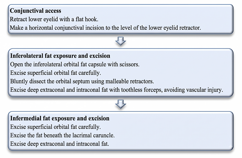
1. Transconjunctival access
The lower eyelid was retracted inferiorly with a flat hook. A horizontal conjunctival incision was made with spring scissors down to the lower eyelid retractor.
This route avoids a cutaneous scar and provides access to the inferior fat compartments. In the authors’ discussion, the transconjunctival approach is positioned as a less invasive alternative to transcutaneous bone-removing procedures, with the potential for less postoperative discomfort and a shorter recovery.
2. Inferolateral compartment decompression
The inferolateral orbital fat capsule was opened with scissors, exposing the superficial fat. A small amount of superficial fat was gently excised while preserving the capsule.
The surgeon then used malleable retractors for blunt dissection through the orbital septum to reach the deeper inferolateral fat. Deep extraconal and intraconal fat was removed with toothless forceps. The authors explicitly warn against excessive resection and vascular injury.
The distinction between superficial and deep fat is crucial. Superficial excision was intentionally conservative; most decompression came from the deep compartments. This approach was intended to recess the globe without producing an over-hollowed lower eyelid.
3. Protect the inferior rectus region
Fat near the inferior rectus compartment was removed cautiously. The stated concern was postoperative inferior displacement of the globe.
For surgeons evaluating this method, that warning deserves emphasis. The goal is not maximal volume removal. It is controlled decompression that preserves globe support and ocular alignment.
4. Inferomedial compartment decompression
The same sequence was repeated medially. After limited superficial excision, the surgeon removed fat beneath the lacrimal caruncle, followed by deep inferomedial extraconal and intraconal fat.
In later cases, the authors selectively added superomedial fat removal to improve balance and achieve a more natural recession. They describe this as a technical refinement, but acknowledge that its long-term value requires further study.
5. Measure the endpoint and leave the conjunctiva unsutured
Excised fat was blotted to remove blood and packed into the outer barrel of an empty syringe for volumetric measurement. Resection stopped when the predetermined volume was reached.
The conjunctival incision was left unsutured after confirming that no epithelium was trapped, and the tissue was returned to its original position.
What the technique achieved
The mean fat volume removed was 3.0 ± 1.0 mL per orbit: 1.4 ± 0.6 mL inferonasally and 1.6 ± 0.5 mL inferotemporally.
At 3 months, mean Hertel exophthalmometry decreased from 19.0 ± 2.7 mm to 16.7 ± 2.8 mm—a mean recession of 2.3 mm (P < 0.001). The observed dose-response was approximately 0.9 ± 0.7 mm of globe recession for each milliliter of fat removed.
Interpupillary distance also narrowed, from 66.2 ± 3.5 mm to 63.9 ± 3.1 mm (P < 0.001). For every 1 mL removed from each eye, IPD decreased by 0.8 ± 0.4 mm. This finding is relevant because perceived ocular prominence is influenced not only by anterior projection, but also by the relationship between the globes and the surrounding facial framework.
Mean intraocular pressure fell from 16.2 ± 2.9 mm Hg before surgery to 14.0 ± 2.4 mm Hg at 3 months (P < 0.001). Best corrected visual acuity remained stable. Fusion image area, used as a functional measure of binocular single vision, decreased slightly at 1 month but was not significantly different from baseline at 3 months.
These data suggest that the procedure produced both anatomical recession and measurable changes in ocular position without a detected loss of central visual acuity over the study period. They do not, however, establish long-term safety or superiority over other techniques.
Safety signals: reassuring, but not negligible
No patient developed permanent visual loss, infection, retrobulbar hemorrhage, or diplopia in primary gaze during follow-up. Reported events were transient and resolved without intervention:
Transient diplopia: 4 patients (17.4%)
Eye pain: 2 patients (8.7%)
Abnormal cheek sensation: 1 patient (4.3%)
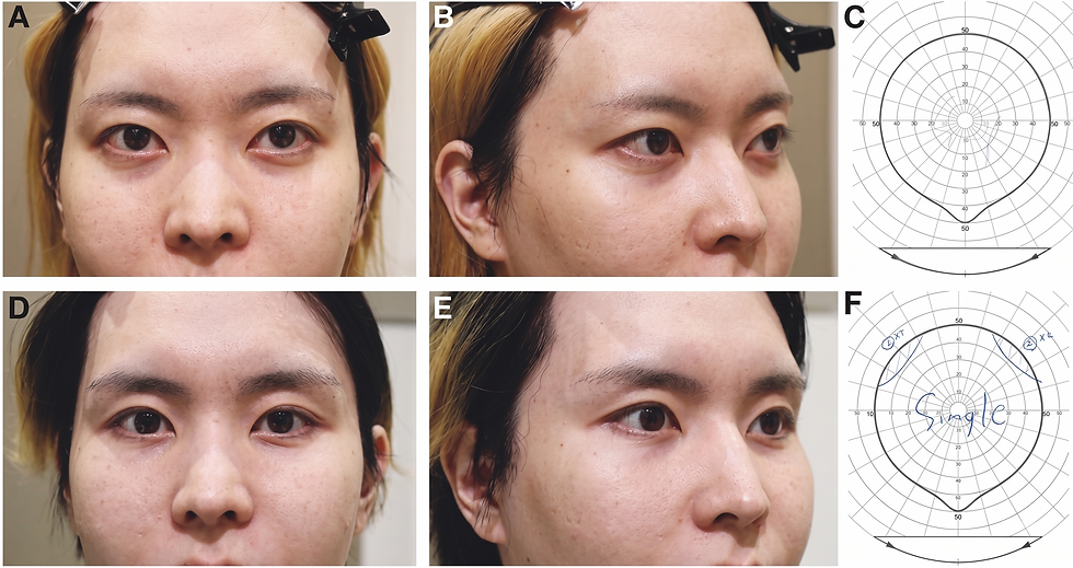
One illustrated patient had slight exotropia in upgaze but no diplopia in primary position. That finding reinforces why alignment should be assessed beyond a simple yes-or-no question about diplopia.
The absence of permanent major complications in 23 patients is encouraging, but it should not be interpreted as proof that the operation is low risk. Deep intraconal dissection occurs near the extraocular muscles, orbital vessels, and optic nerve. The study was performed by experienced orbital surgeons, and its results should be understood in that context.
How should doctors interpret the evidence?
Evidence limitation | Why it matters clinically |
Retrospective, single-center design | Introduces selection and documentation bias and may limit applicability to other clinical settings. |
Small sample of 23 patients | Provides limited power to detect uncommon but serious complications. |
Minimum follow-up of 3 months | Does not establish long-term stability, recurrence, late diplopia, or delayed globe-position changes. |
Procedures performed by six surgeons | Differences in surgical technique and experience may have influenced outcomes. |
Hertel measurements obtained by different examiners | Interobserver variability may have introduced measurement errors of up to 2 mm—substantial relative to the reported mean 2.3-mm reduction. |
No control or comparator group | The study cannot establish superiority over observation, superficial fat removal, or bony decompression. |
No validated patient-reported outcome measure | The study does not quantify patient satisfaction, perceived aesthetic benefit, or quality-of-life improvement. |
The proposed relationship of roughly 1 mm of recession per milliliter of fat is clinically useful as an early planning reference, but the standard deviation was wide. It should not be treated as a guaranteed conversion factor for an individual orbit.

Practical takeaways for orbital and aesthetic surgeons
The paper’s contribution is less about removing a particular number of milliliters and more about where and how that volume is removed.
First, establish the diagnosis. MRI and thyroid testing were integral to excluding TED, inflammation, tumor, and other orbital disease.
Second, plan the endpoint rather than chasing maximal decompression. The desired recession was discussed before surgery and mapped against photographs and MRI.
Third, keep superficial removal conservative. The authors relied primarily on deep inferolateral and inferomedial extraconal and intraconal fat to obtain recession while limiting hollowing.
Fourth, respect the functional anatomy. Avoid vascular injury, exercise particular caution around the inferior rectus compartment, and monitor ocular alignment and binocular single vision after surgery.
Finally, counsel patients using the level of evidence that actually exists. The early results are promising, but the technique remains supported by a small retrospective series with short follow-up. Larger prospective studies, standardized measurements, longer surveillance, and patient-reported aesthetic outcomes are needed before predictability and long-term safety can be firmly established.
The bottom line
Deep orbital fat decompression may expand the surgical options for carefully selected patients with nonthyroidal proptosis who seek globe recession without bone removal. In Yoshida and colleagues’ series, a transconjunctival, deep-fat–dominant technique achieved a mean 2.3-mm reduction in Hertel measurement, narrowed IPD, and lowered IOP, while visual acuity remained stable at 3 months.
The technique’s appeal lies in its precision: limited superficial excision, targeted deep compartment removal, preplanned volumetric endpoints, and deliberate protection of orbital support and motility structures. Its future will depend on whether larger, prospective studies reproduce these outcomes and clarify the long-term balance between aesthetic benefit and orbital risk.
Reference:
Yoshida K, Kashima T, Kikuchi R. Aesthetic Deep Orbital Fat Decompression for Proptosis of Nonthyroidal Origin: A Case Series of 23 Patients. Plastic and Reconstructive Surgery - Global Open. 2025;13. doi:10.1097/GOX.0000000000007180.
Go beyond the techniques—learn directly from Japan’s leading experts and uncover the insights, principles, and approaches behind their expertise:
IFAAS Mini Fellowship (In-OR Observation)
Asian Blepharoplasty: Japan Edition

16-17 November, 2026 Cadaver Hands-On Masterclass: Upper & Lower Blepharoplasty - Singapore [Register Now]
19-20 December, 2026 - Tokyo, Japan - [Register Now]
More Upcoming Global Events