The Five-Step Medial Epicanthoplasty for Asian Patients
- Aug 9, 2025
- 7 min read
Epicanthus, characterized by the longitudinal curved skin folds covering the medial canthus, is a common anatomical feature predominantly observed in Asian populations. It significantly influences the aesthetic appearance of the eyes by partially concealing the medial canthal angle and lacrimal mound. With an estimated incidence of up to 70% among individuals with single eyelids in Asia, the demand for effective epicanthoplasty techniques is steadily rising among plastic surgeons and aesthetic physicians practicing in this demographic.
Despite a variety of surgical approaches developed over the years—ranging from Z-plasty variants to skin resurfacing—there has been no standardized, simplified protocol that ensures consistent outcomes and ease of mastery. This variability poses challenges, especially for surgeons new to epicanthoplasty, and limits widespread clinical adoption.
Overview of the Five-Step Medial Epicanthoplasty Technique
A recent retrospective clinical study by Jie Chen et al. (2025) proposes and evaluates a standardized five-step medial epicanthoplasty method designed to overcome these challenges. Conducted on 306 patients at the Second Affiliated Hospital of South China University, the procedure demonstrated significant improvements in medial canthal aesthetics, high patient satisfaction, and minimal complications.
Anatomical and Theoretical Foundations of the Technique
Understanding the anatomy and biomechanics involved is essential for successful epicanthus correction:
Tension Release as the Core Principle: Over a hundred epicanthoplasty techniques exist, but clinical research emphasizes that the decisive factor for success is the complete release of vertical skin tension, rather than flap design alone. The five-step method prioritizes precise dissection and resection of tension-inducing structures.
Medial Canthal Fibrous Band (MCFB):Identified through cadaveric and clinical studies, the MCFB consists of thickened orbicularis oculi muscle fibers closely adhered to the medial canthal ligament’s superficial surface. This fibrous band acts as a tension conductor, leading to the characteristic vertical fold. Complete resection of the MCFB is fundamental to the technique’s anatomical success.
Technological Innovations of the Five-Step Technique
Unlike traditional methods which rely on fixed preoperative flap designs (Z-plasty, V-Y flaps, etc.), this technique embraces dynamic intraoperative tension assessment:
After MCFB resection and muscle release, the surgeon performs a traction test to confirm the disappearance of skin folds before deciding the flap type.
This flexibility avoids postoperative complications like scar hyperplasia or shape distortion caused by imbalance of local tension vectors.
The technique’s standardization reduces complexity, shortens the learning curve, and improves surgical reproducibility—making it ideal for surgeons early in their practice.
Step-by-Step Surgical Procedure for Five-Step Medial Epicanthoplasty
Step 1: Preoperative Design and Marking
![Source: Chen J, Zhang J, Xi W, Chen W, Yang F. The five-step medial epicanthoplasty: simple and standardized. [BMC Ophthalmol. 2025;]. PMID: 40375074.](https://static.wixstatic.com/media/f6412e_34398613416d494d8df2e2540d1bb385~mv2.png/v1/fill/w_787,h_385,al_c,q_85,enc_avif,quality_auto/f6412e_34398613416d494d8df2e2540d1bb385~mv2.png)
Position the patient supine with eyes gently closed.
Using methylene blue or another fine marking pen, identify three critical points:
Point A: The ideal new medial canthus angle. Determine this by gently stretching the skin medially towards the nose to reveal where the medial canthus angle would naturally fall if the epicanthus fold were corrected.
Point B: The intersection where the free edge of the epicanthal fold meets the upper eyelid margin.
Point C: The extension point along the line from Point B to Point A′ (original medial canthus position before stretching), where the length BC roughly equals length AB.
Importantly, no full flap design is pre-determined at this stage; instead, focus is on the planned area of skin separation (the “ABC flap”).
Step 2: Adequate Subcutaneous Separation
![Source: Chen J, Zhang J, Xi W, Chen W, Yang F. The five-step medial epicanthoplasty: simple and standardized. [BMC Ophthalmol. 2025;]. PMID: 40375074.](https://static.wixstatic.com/media/f6412e_9056ff741cd540d897a513e1f1a620b7~mv2.png/v1/fill/w_787,h_383,al_c,q_85,enc_avif,quality_auto/f6412e_9056ff741cd540d897a513e1f1a620b7~mv2.png)
Using a No. 11 scalpel blade, incise precisely along the lines connecting points A-B and B-C.
With sharp scissors, carefully elevate the skin flap ABC, maintaining a thickness that preserves subdermal vascularity but allows adequate exposure of the underlying orbicularis oculi muscle and connective tissues.
The dissection zone should be about 10 mm long and 8 mm wide, creating a partial 270° circular flap.
Thoroughly release attachments of the anterior orbicularis oculi muscle from the medial canthal skin to free the skin flap.
Take care to preserve the superficial vascular network to minimize bleeding and enhance healing.
This step aims to completely disconnect the dense connective tissue bridging skin and muscle, which transmits abnormal tension causing the epicanthal fold.
Step 3: Skeletonization of the Medial Canthal Ligament Surface
![Source: Chen J, Zhang J, Xi W, Chen W, Yang F. The five-step medial epicanthoplasty: simple and standardized. [BMC Ophthalmol. 2025;]. PMID: 40375074.](https://static.wixstatic.com/media/f6412e_ba668cef22434b5ca3a5af90b09ee9ac~mv2.png/v1/fill/w_787,h_383,al_c,q_85,enc_avif,quality_auto/f6412e_ba668cef22434b5ca3a5af90b09ee9ac~mv2.png)
Continue blunt and sharp dissection to remove fibrous connective tissue adherent to the superficial surface of the medial canthal ligament.
Expose the white medial canthal ligament extending horizontally from the medial canthus angle toward the nasal dorsum deep fascia.
Fully release heterogeneous and misaligned orbicularis oculi muscle fibers that tether to the medial canthal ligament or surrounding soft tissue.
Sever muscular fibers bridging the orbicularis oculi and the epicanthal fold to eliminate vertical and horizontal traction forces.
The goal is to “skeletonize” the ligament, freeing it completely from overlying muscle and connective tissue without damaging ligament integrity.
This extensive release mitigates both vertical skin tension and horizontal pull, which are biomechanical contributors to epicanthus formation.
Step 4: Medial Canthal Ligament Tension Reduction
Using a 6-0 polydioxanone (PDS) absorbable suture, perform a zigzag suture technique directly on the medial canthal ligament.
The zigzag pattern distributes and reduces tension more evenly than simple linear sutures.
Concurrently, consider suspending the nasal subcutaneous tissue or deep nasal fascia to adjacent fixed structures to improve skin flap fitting and further reduce tension.
Throughout this step, continuously perform intraoperative traction tests by pulling the medial canthal skin towards the nasal side and observing the disappearance of the fold. This real-time assessment guides the adequacy of tension release.
Carefully adjust suture tension to balance sufficient skin apposition without over-tightening, which could cause distortion or ischemia.
Remember that tissue shrinkage and remodeling will occur during healing, so slightly over-correct tension reduction if needed.
Step 5: Skin Management and Closure
After tension reduction, gently smooth the skin flap to assess redundancy or defects.
If excess skin is present, mark and excise carefully to prevent dog-ear formation or unnecessary skin tension.
Skin closure options depend on intraoperative flap geometry:
Y-V advancement flap: Useful when mild skin advancement is needed without creating large defects.
Z-plasty or modified Z-flap: Ideal for redistributing tension lines and breaking up linear scars, especially when a triangular skin defect exists below the medial canthus angle.
The freed ABC flap may be repositioned into the defect site to achieve optimal contour and tension.
Use a 6-0 absorbable suture for subcutaneous closure to minimize tension on the skin edges.
For skin closure, employ a 7-0 fast-absorbing collagen suture or equivalent fine suture material to promote minimal scarring and excellent cosmesis.
Apply sterile gauze dressing and initiate cold compresses immediately post-op to reduce edema and hematoma risk.
Replace wound dressing on postoperative day 2 and instruct the patient on scar management protocols (silicone gel, sun protection).
Additional Technical Tips and Pearls
Maintain meticulous hemostasis throughout to avoid hematoma, which can compromise healing and increase scar risk.
Minimize cautery use near skin edges to preserve skin viability.
Postoperative scar management is critical, particularly in patients prone to hypertrophic scarring or those with darker skin tones.
Counsel patients preoperatively on realistic expectations regarding scar color changes, especially in those with under-eye dark circles or pigmentation differences.
For cases with preoperative severe medial canthal ligament laxity, anticipate greater intraoperative adjustment of tension sutures and skin flap design.
In the presence of mild asymmetry or skin irregularities postoperatively, minor touch-up procedures may be planned after 6–12 months.
This 5-step medial epicanthoplasty approach ensures controlled, reproducible correction of the epicanthal fold by addressing the key anatomical and biomechanical factors underlying the deformity. The combination of careful tissue release, dynamic tension assessment, and adaptable skin flap management underpins the high success and satisfaction rates reported in clinical practice.
Results
The five-step medial epicanthoplasty technique was applied in a cohort of 306 patients, predominantly female (295 females, 11 males), with an average follow-up of 14.2 months. The procedure demonstrated statistically significant improvements in key anatomical parameters and high patient satisfaction:
Palpebral Fissure Length (PFL): Increased on average by 14.9%, indicating effective enlargement of the visible eyelid aperture.
Intercanthal Distance (ICD): Reduced by an average of 8.6%, reflecting successful medial canthus repositioning and fold correction.
Medial Canthal Angle and Canthal Tilt: Both angles showed significant decreases postoperatively, contributing to a more natural and aesthetically pleasing eye shape.
![Source: Chen J, Zhang J, Xi W, Chen W, Yang F. The five-step medial epicanthoplasty: simple and standardized. [BMC Ophthalmol. 2025;]. PMID: 40375074.](https://static.wixstatic.com/media/f6412e_d6abf8c8ab614e1899884f22187b9169~mv2.png/v1/fill/w_787,h_788,al_c,q_90,enc_avif,quality_auto/f6412e_d6abf8c8ab614e1899884f22187b9169~mv2.png)
Image: a) Before surgery, b) postoperative day 7, c) postoperative month 6
Complication rates were low and manageable:
Minor postoperative hematoma and swelling occurred in only 2 cases, resolved with prompt intervention.
Scar hypertrophy developed in 85 cases but was effectively treated with silicone gel, steroid injections, and laser therapy, with scars resolving within 12 months.
Recurrence occurred in just 2 cases, and asymmetry requiring revision was seen in 4 cases.
Overall, over 95% of patients reported satisfaction with the surgery outcome, with 96.4% willing to recommend the procedure to others.
Photographic follow-ups demonstrated minimal and well-concealed scarring, with most patients showing fine scars without retraction at long-term follow-ups up to 3 years.
These results affirm that the five-step epicanthoplasty method is not only technically reproducible and beginner-friendly but also effective in achieving stable aesthetic improvements with minimal complications.
Limitations and Future Directions
The technique is less effective for inverted epicanthus, which involves abnormal lower eyelid tension and soft tissue misalignment requiring specialized approaches.
No control group or randomized comparisons were included in the current study, limiting direct efficacy comparisons against traditional methods.
Future multicenter randomized controlled trials comparing operative time, scar incidence, recurrence, and long-term patient satisfaction are needed to further validate this technique’s benefits.
Clinical Advantages for Your Practice
Shortened Learning Curve: Junior surgeons mastered this technique within days, performing independently after limited supervised cases—compared to months with complex traditional methods.
Reduced Complications: Standardization and dynamic tension management decrease risks of hypertrophic scars, asymmetry, and recurrence.
High Patient Satisfaction: Over 95% effectiveness rate with excellent aesthetic results and minimal visible scarring.
Conclusion
The five-step medial epicanthoplasty is a simple, standardized, and reliable technique for correcting epicanthal folds in Asian patients. Its focus on comprehensive tension release, anatomical precision, and dynamic intraoperative decision-making leads to consistent, reproducible results that are easy for beginners to master. This method holds significant promise for wider clinical adoption and improved patient outcomes in epicanthoplasty.
Reference:
Chen J, Zhang J, Xi W, Chen W, Yang F. The five-step medial epicanthoplasty: simple and standardized. [BMC Ophthalmol. 2025;]. PMID: 40375074.
Learn techniques behind Upper & Lower Blepharoplasty
in our upcoming Master Class:
IFAAS Fresh Cadaver Master Class (Hands-On)
Upper & Lower Blepharoplasty Level 1: Suture & Minimally Invasive Techniques
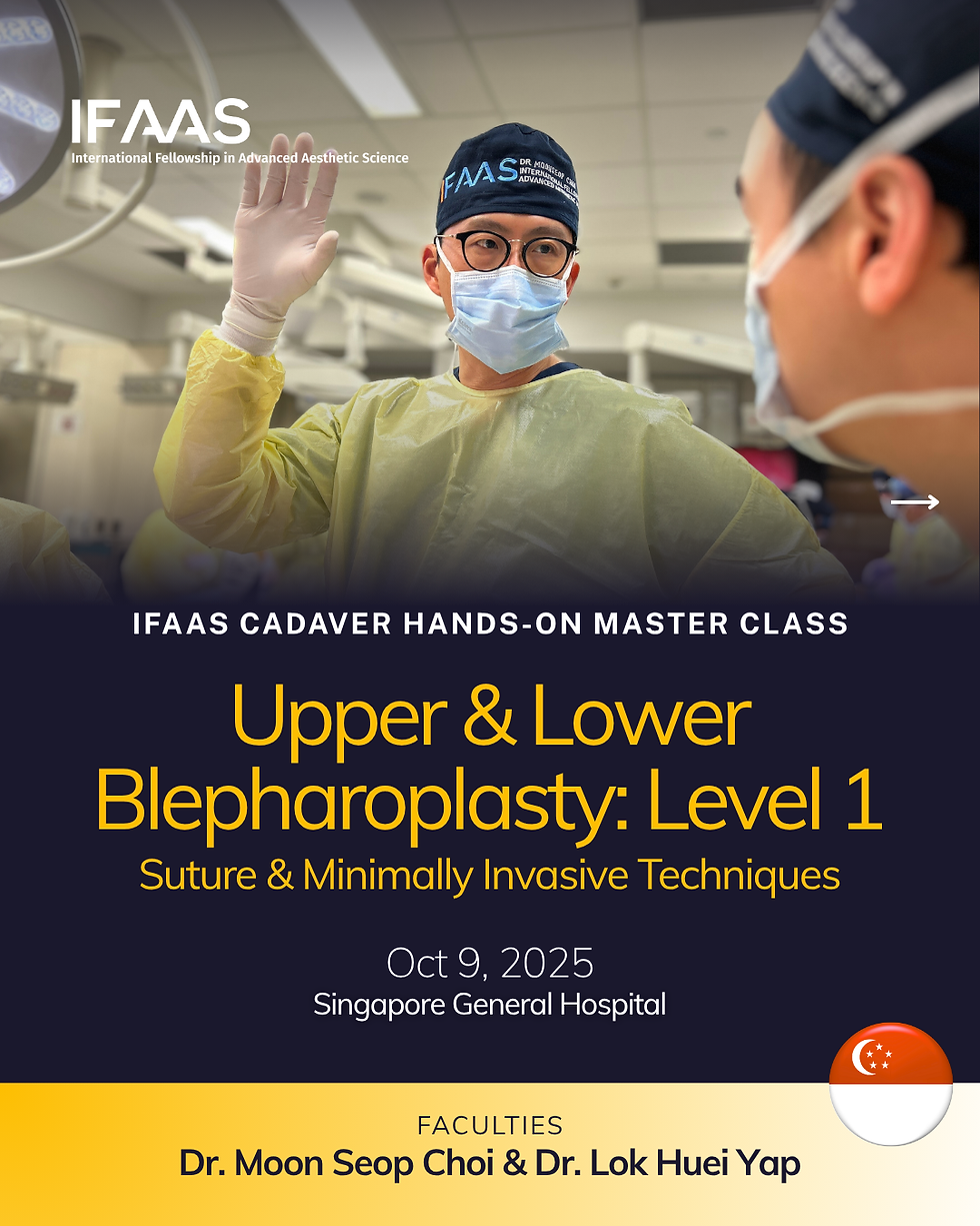
Oct 9, 2025 - Singapore General Hospital - [Register Now]
Oct 10, 2025 - Singapore General Hospital - [Register Now]
Dec 13-14, 2025 - Tokyo, Japan - [Register Now]
More Upcoming Global Events
Comments